Do X Rays Show Muscles
The best imaging study is MRI which shows muscles the best. Doctors use X-rays of the shoulder to examine the bones and determine if any bone spurs are present or if there is a fracture which can cause similar symptoms.
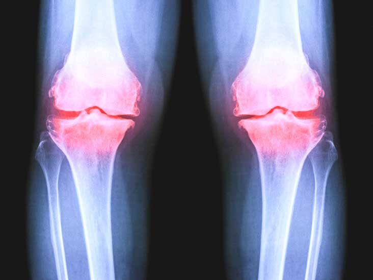
Osteoarthritis Of The Knee X Ray
MRIs work differently and visualise tissues containing water or lipids.
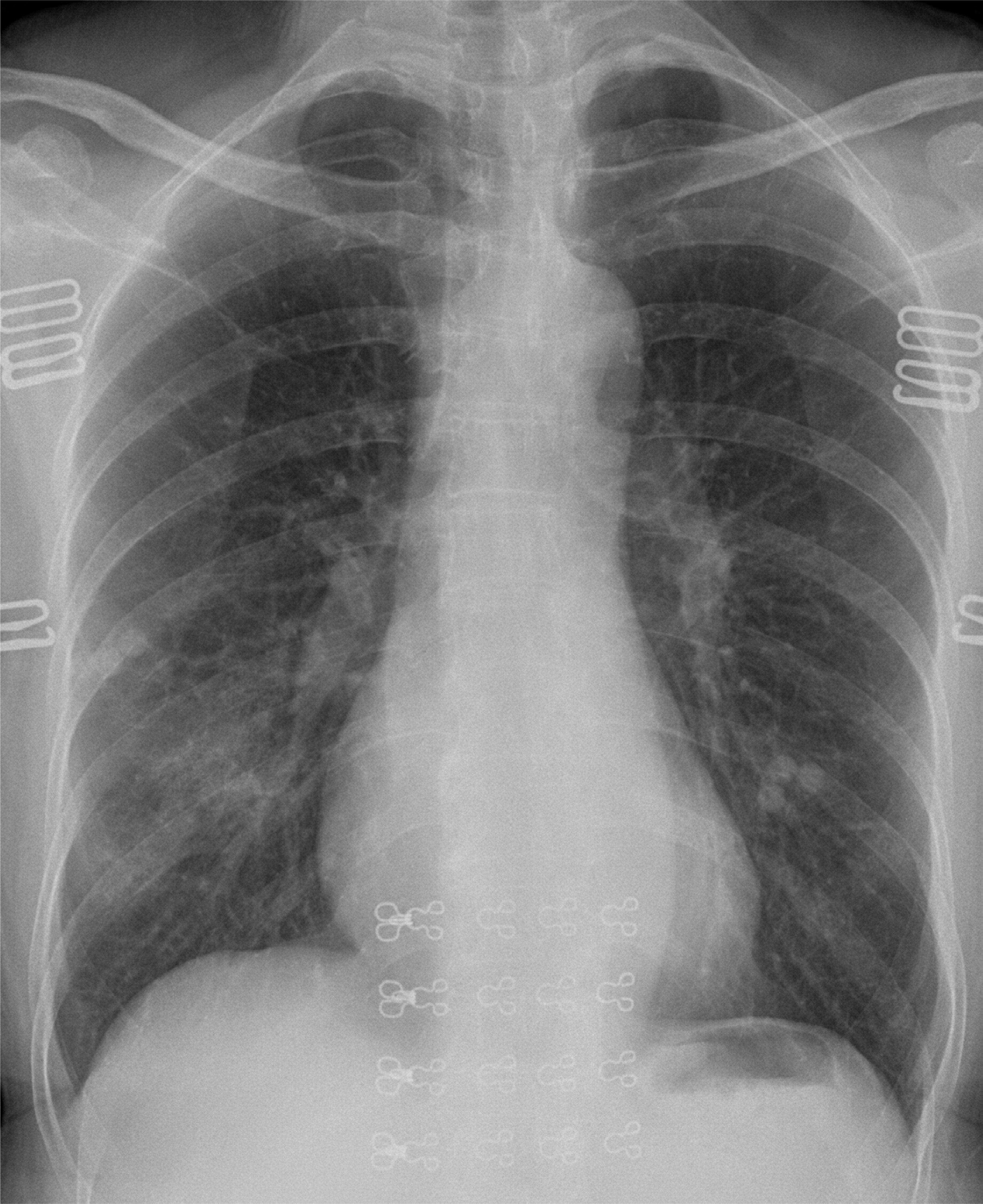
Do x rays show muscles. They dont show soft tissue structures like the tendons ligaments meniscus in the knee muscles cartilage etc. X-rays X-rays use high-energy beams of light to create pictures of the body. Depending on which.
X-rays are often called the workhorses of the radiology department and this type of spinal imaging test best detects bony structural problems. Doctors may order a CT scan when they need greater detail regarding bones muscles or both. The machine sends a beam of radiation through your body.
X-rays do not show soft tissues such as muscles bursae ligaments tendons or nerves. However you can see muscles on a CT scan much better. X-rays can also help doctors rule out osteoarthritis of the shoulder as the cause of your symptoms.
Sensory nerves then communicate from the skin and muscles back to your brain and spinal cord causing you to feel pain. These nerves can all be damaged by sports injuries falls or accidents. Ultrasounds can be used to monitor the muscle and.
Damaged nerves cannot be seen on a regular X-ray. X rays are barely blocked by soft tissues so only faint shadows are left on the sensor or film. To answer that I need to explain why many people dont feel like they are necessary.
A plate behind the body part captures the variations of the energy beam after it passes through skin bone muscle. An ultrasound uses sound waves to show pictures of your muscles and tissues on a monitor. Muscles that spasm are a functional aspect of lower back disorders and cannot be seen on an MRI or X-ray unless there is an antalgic list which would be evident on standing X-rays.
Your hard dense bones block that beam so they show up as white on the film below you. Doctors at NYU Langone often use ultrasound to diagnose muscle tendon and ligament injuries. This is because ultrasound uses high frequency sound waves to produce an often clearer picture of soft tissue such as muscles and ligaments compared with X-ray images.
Regular x-rays may show indirect evidence suggestive of a possible tendon or ligament injury eg. Victor Scarmato Radiologist East Meadow NY. In standard X-rays a beam of energy is aimed at the body part being studied.
Muscle spasms are typically a result of back disorders and not a cause. To help determine whether the joint has been damaged by injury a doctor may use an ordinary non-stress x-ray or one taken with the joint under stress caused by certain positions stress x-ray. X-rays wont show a torn rotator cuff but can rule out other causes of pain such as bone spurs.
You can see muscles on radiographs x rays are what make the Radiograph. Sometimes X-rays can show evidence that a muscle might be injured but you really need a much more sophisticated imaging study to see muscle damage. Although soft tissues such as muscles tendons and ligaments do not show up on X-ray doctors often order X-rays when you have back or neck pain to rule out the possibility that a spinal fracture tumor or degenerative joint disease could be causing or contributing to your pain.
It is used to check for muscle injuries broken bones and damaged blood vessels. There are several ways of looking inside the body including X-rays computed tomography CT scans and magnetic resonance imaging MRI. Motor nerves on the other hand control voluntary movement and actions by communicating to muscles from the brain and spinal cord.
For example muscles and ligaments do not show up very well on an X-ray scan. A CT scan works like a regular x-ray except an x-ray beam will move in a circle around your body. They can be seen on CAT scan or MRI and in fact MRI is recommended for examining details.
Another one high-resolution ultrasound offers some advantages over the others for musculoskeletal problems. What can I do to help a muscle strain heal. X-rays are helpful for information about bones but may not provide enough information about muscles.
But high energy X-rays are used to visualize bones. A x-ray machine uses a computer to take pictures of your arms legs back or abdomen. As you may or may not know xrays just show bones.
While X-rays show irregularities they are very limited in what they are able to display. Fluid but do not directly show ligaments or tendons and are best for evaluating the bones and for fracturesAn MRI would be the best imaging study to more directly assess for ligament and tendon injury. Many lower back pain cases improve in days or a few weeks.
Ultrasound scans are quick and painless.

Tests For Musculoskeletal Disorders Bone Joint And Muscle Disorders Merck Manuals Consumer Version
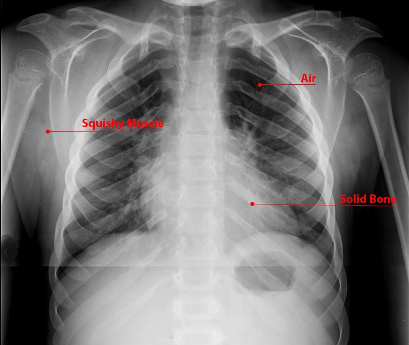
What Is An X Ray For Kids Radiology And Medical Imaging

Do I Really Need An X Ray Or Mri For Lower Back Pain
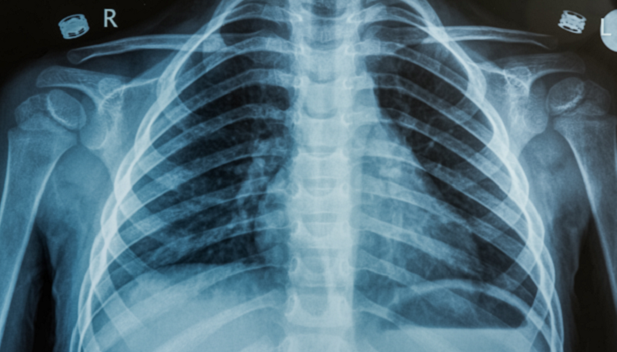
Lymphoma Action Scans X Ray Ct Pet And Mri
X Rays Undergraduate Diagnostic Imaging Fundamentals
Abdominal X Ray Johns Hopkins Medicine

Fractures X Ray Stanford Health Care

Ai Can Assist In Triaging Abnormal Chest X Rays Physics World
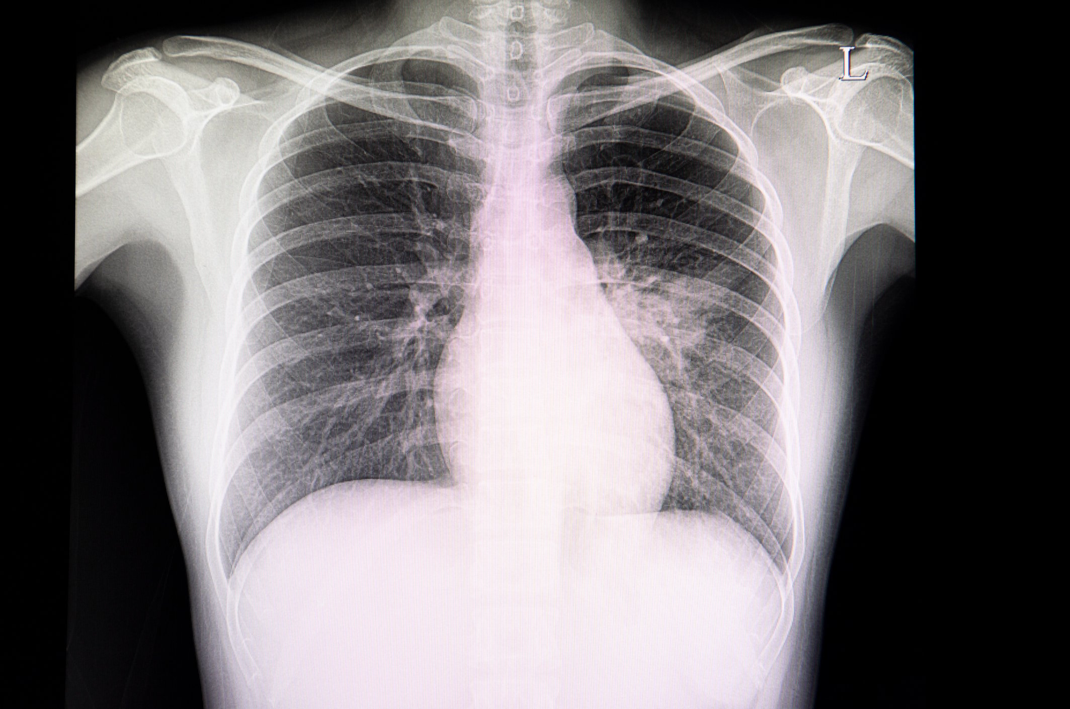
New Research Finds Chest X Ray Not Reliable Diagnostic Tool For Covid 19 Imaging Technology News

Tests For Musculoskeletal Disorders Bone Joint And Muscle Disorders Merck Manuals Consumer Version

Chest Imaging What Your Chest Scan Can Reveal Touchstone Imaging Centers

Chest X Rays Show More Severe Covid 19 In Non White Patients Imaging Technology News
Snapping Scapula Syndrome Pictorial Essay
X Rays Of The Extremities Johns Hopkins Medicine
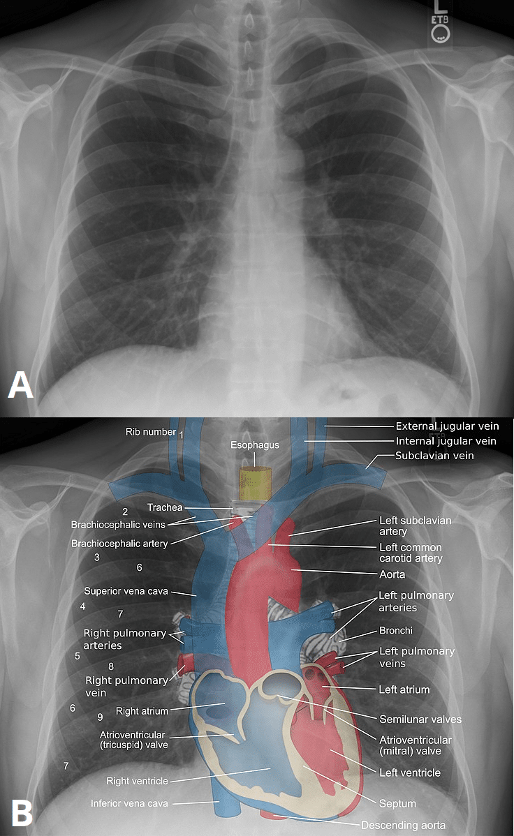
Plain Film X Ray Principles Interpretation Teachmeanatomy
:background_color(FFFFFF):format(jpeg)/images/library/12296/chest_PA.jpg)
Radiological Anatomy X Ray Ct Mri Kenhub


Post a Comment for "Do X Rays Show Muscles"